


Osteochondrosis is a chronic disease that is symptomatic expressed with dystrophic disorders in the joint cartilage. Most often it occurs in the osteocondrosis of the spine, when changes occur in discs between the vertebrae and the intervertebral joints. Depending on the localization, the cervix, chest and lumbar osteochondrosis can be distinguished. Osteochondrosis is also often found in which all spinal parts suffer. Pathology requires consultation with the doctor and an integrated approach to treatment.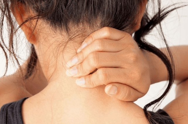
Description
Spinal diseases, like other chronic diseases, quickly become "younger". If the back and joint pain were previously disturbed by the elderly, patients aged 18-30 are increasingly treating doctors.
Scientists consider the immediacy of a person as a prerequisite for the development of the disease, and osteocondrosis is also facilitated by a longer seating situation. In addition, besides age, joint cartilage loses elasticity and elasticity, thin; Intervertebral discs lose moisture and absorbing capacity, and become vulnerable at the time of physical effort.
Reasons
The causes of the cervical osteochondrosis are in load and influence the cervical region under different conditions. Accordingly, the cervical muscles begin to decrease intensely, thus compensating for this load, which resulted in cramps and blood flow violations in the area.
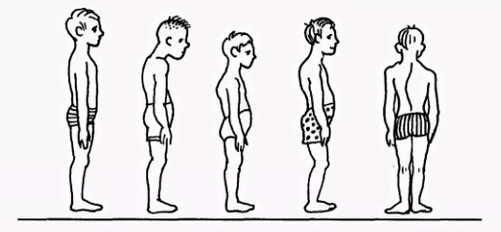
- Violations of posture scholiosis, bending, round back, kyphosis and other posture, even if insignificant, cause severe violation of the spinal column balance. As a result, the load on the intervertebral plates is unevenly distributed, provoking deformation and increased wear. The vertebrae get closer, causing violations of nerve processes, and the cervical osteochondrosis develops quite quickly. Similar consequences have a violation of posture caused by a change in the natural position of the ribs.
- Muscle cramps cramped reactions to the back muscles, the breast, the press, the press can lead to the fact that parts of the body are very tense. As a result, the general equilibrium position of the body is disturbed, which causes change in the spine. Deformations can affect the region of the cervix or other parts of the spinal column, causing osteocondrosis of the chest, cervix and lumbar parts.
- Violation of blood supply, as vertebrates have no direct contact with the circulatory system, receive food from the surrounding tissues. Violation of blood supply to the cervical spine leads to the fact that discs do not receive sufficient fluid for rehydration (restoration of form due to moisture absorption) and renewal of cartilage tissue. As a result, their wear accelerates and the distance between the vertebrae of the cervix is reduced, leading to osteochondrosis.
- Involvement violation, a decrease in nerve roots sensitivity leads to abnormal changes in their structure, as a result of which the displacement and deformation of the vertebrae of the cervix remains unnoticed. After all, sensitivity disorders lack pain.

- Diseases of the internal organs lead to a violation of the general balance of the body due to the improper position of the internal organs, the displacement and the reduction of various dysfunctions. As a result, this acute affects the position of the spinal column - the cervix, the lumbar vertebrae move and deform, leading to the appropriate types of osteochondrosis.
In general, the effects of osteochondrosis of the cervical region develop the effects of harmful external factors that violate the natural equilibrium position of the spinal column and other human body systems.
Diagnosis
Diagnosis of cervical osteochondrosis begins with all the necessary information about the patient. The specialist asks the person's complaints, he or she is interested in his professional activities and how he spends the weekend. An important aspect is the presence of osteochondrosis in parents, grandparents, because it is a hereditary disease.
The doctor then leads directly to the patient's visual examination. Study the cervical compartment and back for the curvature of the posture with the cervical region. This allows the specialist to evaluate the degree of disease development, as in advanced cases the palpation of the cervical region causes sharp pain.
You should pay attention to:
- the severity of cervical lordosis;
- the height of the shoulders in the patient;
- the possibility of asymmetry of supraclock areas;
- the possibility of neck asymmetry (such as congenital pathology or sharp muscle spasm);
- The shoulder belt and upper limbs (for example, a state of atrophy of a single -sided muscle may indicate compression of the cervical spine);
- Location of the chin - the chin should be normal along the middle line;
- Neck movement (bending, better, rotation and rotation).
The palpation is done in the initial position of the patient:
- lying on his back;
- lying on the stomach;
- Sitting in a chair.
We also test the amount of movements. This is performed in the initial position of the patient sitting on the chair (for fixing another spine).
Distinguish the following basic movements in the cervix region:
- bending;
- extension;
- slopes to the right and left;
- Rotation.
About half of the bending and the extension volume occur between the back of the head, the C1 and C2 vertebrae. The rest of the movement is carried out due to the underlying vertebrae, with the high movement of the C5-C7 vertebrae. The side inclination is evenly distributed between the vertebrae.
Further studies are prescribed to make the diagnosis accurately:
- X graves of the cervix region. This method can be useless in early stages of the disease, but in advanced forms.
- CT (computer tomography). It allows you to view structural changes in the vertebrae, but it is impossible to determine the size of the hernia between the vertebrae.
- MRI. It is considered the most effective method for diagnosing cervical osteochondrosis. You can determine the size of the hernia between the plates and the extent of their development.
- The doctor can also prescribe a duplex examination that allows you to determine the normal blood circulation of arteries.
Treatment
Treatment of cervical osteochondrosis is a complex therapy that includes taking medicines, gels and various physiotherapy measures. Massage of therapeutic gymnastics and the problematic area play an important role.
Medicine
It is impossible to eliminate the consequences of the degenerative-registrophical changes in the spine without the use of medicines. The use of drugs in the cervical region in the treatment of osteochondrosis is one of the main points in the complex approach to healing used by most traditional medical professionals.
It is designed to solve more problems, including:
- Stop the symptom of pain and remove the inflammatory process;
- Remove the muscle spasm;
- stimulates the regeneration of cartilage and bone cells;
- Confirm the protective properties of the body, increase immunity;
- Improve the general condition by eliminating other symptoms that interfere with healing.
Depending on these goals, all medicines prescribed by a physician with osteochondrosis in the cervix can be conditionally divided into the following groups:
- Analgesics (non -sertoid drugs that relieve pain).
- Anti -inflammatory (steroid) hormonal drugs that alleviate inflammatory phenomena and thus eliminate pain.
- Condroprotectors are drugs that contain substances that replace cartilage components - chondroitin, hyaluronic acid.
- Musorelaxants. These are drugs that relax in muscle tone. It is used in surgery and orthopedia as an complementary drug to stop pain. Such drugs are administered by Parentel and therefore always under the supervision of a doctor.

- Vitamins. Vitamins are prescribed with osteochondrosis of the cervix, which have a beneficial effect on the peripheral nervous system and improve conductivity. Water -solving vitamins: B1, B6, B12, Fat Solving Vitamins: A, C, D, E. In recent years, medicines containing analgesics and vitamin components have been combined.
- Ointment and gels for external use.
Physiotherapy
The main purpose of physiotherapy treatment is to stimulate the regeneration process and eliminate pain. The most popular methods for treating cervical chondrosis are as follows:
- Ultrasound. Physium with ultrasound waves is used to alleviate severe pain and inflammatory reaction. Ultrasound is the massage of the neck tissue, after which the metabolism is activated.
- Vibration massage. During vibration massage, the effect of pain on the area of pain is due to mechanical oscillation movements. For the right behavior of this physiotherapy, a tape vibrator is usually used.
- Electrophoresis. This method of physiotherapeutic supply of osteochondrosis of the cervix is carried out using diadynamic and modulated currents and electrical fields when the body is administered into the tissues of the drugs. Electrophoresis perfectly relieves cramped syndromes and eliminates the pain of inflamed muscles.
- Magnetic therapy. The essence of magnetic therapy is explained as physiotizing in the osteochondrosis of the cervical region by using the magnetic, the different sizes, using constant or variables. This method can help the patient to remove pain and stop the inflammatory process of the fireplace. The procedure is often performed at home after acquiring a special magnetic device.
- Dutoensor therapy. Currently, it is a rather popular physiotherapy method that consists of stretching the spinal column under the weight of the patient's body. A specially arranged mattress is required to perform such a procedure, which has oblique ribs and changes the place under the weight of their own body. Muscle tone is normalized, leading to relaxation.
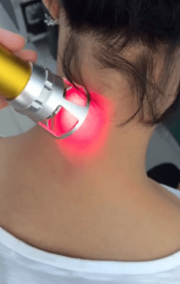
- Laser therapy. The laser has a complex effect on the focus of inflammation, activating biological processes in the tissues of the nervous system. This allows you to have a positive effect from treatment. The complex effect on the body consists of anti -inflammatory, analgesic and wound healing effects. One laser treatment procedure should not exceed 15 minutes. This is the optimal time for the Helic-Neon laser exposure in the affected areas. In this case, the duration of the laser should not exceed 2 minutes for a pain.
- Balneotherapy. The benefits of mineral water have long been known, the basis of balneotherapy. The procedure means the active use of water resources in the treatment of osteochondrosis. In addition to accepting baths, various souls and active swimming in the pool, therapy involves the use of therapeutic mud in painful areas of the body. The therapeutic effect is the concomitant effect of chemically active substances in the water under different temperature conditions. The technique allows you to stop the pain syndrome by improving the local microcirculation of the tissues.
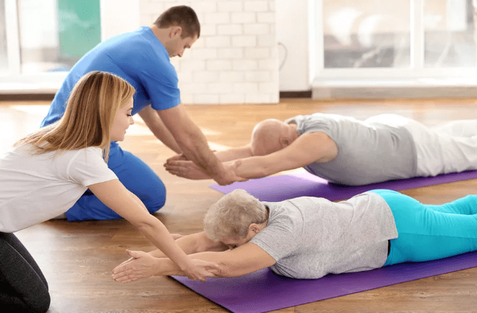
Practice therapy
It should be remembered that exercise therapy is not performed when signs of aggravation begin: pain. The LFK can increase and cause discomfort after the LFK complex.
There are many general recommendations for charging:
- Physical education must do indoor ventilation, a great opportunity on the street.
- Classes are only performed during the remission of the disease (if there is no symptom).
- There is an alleged, non -disturbing movement and breathing of exercise therapy.
- All movement is smooth, the amplitude and the number of repetitions gradually increase.
- If the pain begins, you should stop the lesson immediately.
- Prevents classes and complete the pressure and pulse measurements. When these indicators differ from normal, the load should be reduced.
- We recommend that you listen to your breath during the lessons, which increases your efficiency. All stretching exercises are exhaled.
- It is very important to gradually increase the load and the number of repetitions, which reduces the risk of injury and prevents overtime.
- Exercises are important for regular implementation, so you can achieve quick results.
- Before starting independent classes, you should consult a doctor and agree with a practical set.
Recommended exercises in the starting position on the stomach:
- The head is on the forehead at the end, his hands on the back of the head, parallel to the floor with the floor. Raise your head from the floor, keep this position to 4 accounts, inferior and relax. Repeat 2-4 times.
- The head is at the chin stop, the palm under the chin. Over time, stretch your arms forward, two - to a widespread side, three - stretches forward, four - the starting position. Repeat 2-4 times.
- The hands stretched out in advance. Swimming the "Rabbit" style, 4-8 times repeated.
- The palm under the chin, emphasizing the forehead palm. Alternatively, remove the bottom of the bottom. Repeat 4-8 times.
Recommended exercises in the starting position on the side (on the right, on the left):
- The right hand is extended, the right ear lies on it, lift your right hand with your head, hold the position to 4 accounts, lower and relax. Repeat 2-4 times.
- The left hand rests on the floor in front of the chest, the left foot moves the movement of the fly forward. Repeat 6-8 times.
- The left side along the body, lift your left hand up and out, bottom, bottom. Repeat 2-4 times.
- The left hand on the thigh. Pull both knees on the chest when exhaled, straighten your leg with inspiration. Repeat the exercises 2-4 times.